Endobronchial Ultrasound Bronchoscopy (EBUS) Procedure
April 25, 2022

What Is EBUS Bronchoscopy?
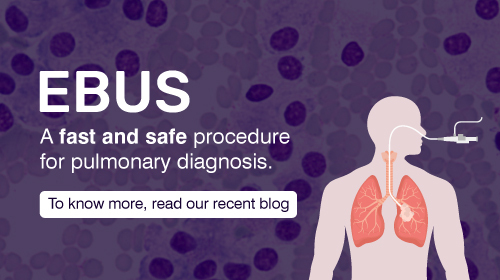
The moment when we hear the word “EBUS”, we tend to naturally misinterpret it as something related to travel and transport. But, the word EBUS in Pulmonology refers to Endobronchial Ultrasound, a minimally invasive bronchoscopic procedure performed by Pulmonologists which provides a real-time ultrasound imaging of the lung and mediastinum and helps to obtain tissue via Transbronchial needle aspirations (TBNA), fine needle biopsies (FNB) and core biopsy for diagnosing lung cancer and stage them to assess operability, re-diagnosis & staging in case of progressive lung and other metastatic tumours, inflammatory conditions, chronic granulomatous infections and rare autoimmune conditions.
Just like any other diagnostic tool such as Endoscopy, Colonoscopy or Coronary Angiogram, EBUS-TBNA is a semi-invasive procedure performed by Interventional Pulmonologists by manoeuvring the flexible EBUS Scope through the mouth/ nasal cavity into the trachea (windpipe) to visualize the lesion using low frequency (5-15Hz) real time ultrasound probe with great accuracy. It further enables to perform a FNA or biopsy without obvious pain or any other major complications that we may usually encounter in mediastinoscopy or other invasive thoracic procedures.
What is TBNA?
Before the invention of EBUS, Conventional TBNAs (also referred to as blind TBNA in layman terms) were performed in the later half of the 20th century which had variable diagnostic accuracy of 40-75% and resulted in more than a few complications. It also resulted in false negative reports which has more often led to a complicated path. But, after the revolutionary invention of EBUS by Yasufuku and colleagues, Japan in 2004, it has slowly but steadily become the diagnostic modality of choice for sampling the mediastinum and peribronchial areas of the lung and has a diagnostic accuracy of > 90% for detection of lung cancer, more than 95% in tuberculosis and other granulomatous infections and approximately 80-85% in sarcoidosis.
What can EBUS Diagnose?
In the case of Mediastinal lesions (masses and nodes), symptoms are non-specific and vague and even may be completely asymptomatic (nearly 40-60%) and identified on incidental imaging (e.g. a CT Chest as part of master health check-up/ insurance/ job fitness). But if a person has symptoms such as cough with or without sputum production, breathing difficulty, chest pain, blood in sputum, difficulty in swallowing, recent change in voice, loss of weight and appetite, chest discomfort, wheezing and noisy breathing they often point towards some underlying disease. In an era of precision and personalized medicine where “tissue is the issue” a confirmatory diagnosis to the varied etiology with utmost accuracy is possible with EBUS, thereby it helps to overcome the diagnostic dilemma leading to undue delay in appropriate treatment as well as unwarranted treatment.
EBUS is not only known for its accuracy but also for its safety. It is extremely safe (99.3%) with minimal complications such as minor bleeding, cough, fever and in rare cases mediastinitis. Recently Pulmonologists especially in the last decade have started to use the EBUS Scope in the esophagus and procure samples from mediastinum, a technique popularly referred to as “EUS-B-FNA” in the field of Pulmonology. It is of use especially in high-risk pulmonary patients who may not tolerate a bronchoscopy and is associated with minimal complications.
What is EBUS TBNA Test?
EBUS-TBNA is a day care procedure and it takes approximately 30-60 minutes depending on the indication. Even if we were to include the recovery and observation time it does not usually take more than 4 hours. Unlike major surgeries or other complicated procedures, the patient may not be subjected to general anaesthesia but can be performed under ‘conscious sedation’ (a term used by the British Society of Anaesthesiologists refers to a sedation where verbal contact with patient is possible throughout the procedure) making a sophisticated procedure simplified for both patients and the bronchoscopist.
Besides, there are only a few and far-fetched centres with this expertise in our city and only handful in the entire state with a fully functional set-up, including ours, thereby, enabling to unlock the door to lung and mediastinum lesions which were treated empirically since time immemorial and even in this modern age without any obvious cytological or histopathological confirmation. Yet, there seems to be an appalling paucity of awareness about EBUS among our patients and physicians alike as well as the general public. I conclude this message with a small quote, “Knowledge is Wealth” & We at the Department of Pulmonary Medicine, Rela multispeciality hospital have the luxury of EBUS with us and I have put an earnest effort to provide a glimpse of this modality through my letter, which has changed the landscape of Interventional Pulmonology across the Globe, something akin to coronary angiogram (CAG) in Cardiology.


