Electrocardiogram vs. Echocardiogram
March 18, 2024

Electrocardiogram and Echocardiogram
Diagnostic testing may be necessary if you experience odd symptoms, such as palpitations or shortness of breath, that could be related to your heart.Electrocardiograms and echocardiograms are two of the most frequently performed tests.Heart-related problems can be diagnosed with noninvasive diagnostic techniques such as an echocardiography and electrocardiogram.
Contents
- What is an Electrocardiogram (ECG)?
- Types of electrocardiograms
- What is an Echocardiogram (ECHO Test)?
- Types of echocardiograms
- Comparing ECG vs. ECHO Test Results
- Requirement for ECG vs. ECHO Test
- Procedural differences between an ECG and an ECHO Test
- Procedural duration for ECG vs. ECHO Test
- Details provided by ECG vs. ECHO Test
- Conclusion
- Frequently Asked Questions
Electrocardiogram(ECG)
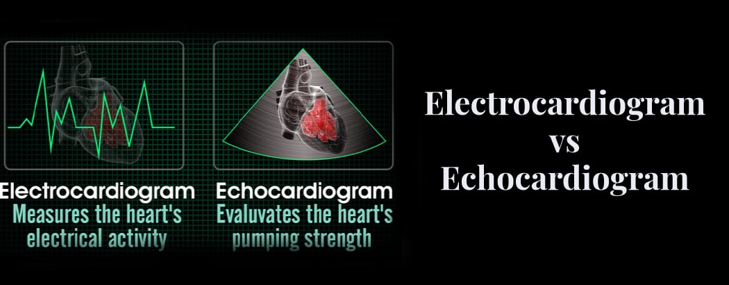
An electrocardiogram (ECG) is a non-invasive and painless device used by doctors to measure the electrical activity of the heart. Every time the heartbeats, it produces an electrical impulse that facilitates the pumpsing of blood through the heart. Doctors use ECGs to determine whether the heart’s electrical activity is normal or not.
Wave patterns representing your heart’s electrical rhythm are displayed on an ECG. Unpredictable, erratic, or non-standard waves may indicate heart problems. An ECG is used to detect irregularities in your heart and to assist your doctor understand how well your heart is functioning.
Types of Electrocardiograms
The different types of electrocardiograms are:
Exercise ECG: During an Exercise ECG, a person is made to exercise while their heart activity is monitored.
Holter Monitor: An ECG is worn for a predetermined amount of time. The gadget can be worn around the neck or fastened around the waist like a belt. The electrodes will be affixed to the skin by the technician and connected to the recording device.
Echocardiogram(ECHO Test)
Echocardiography is a medical test that uses sound waves to create an image of your heart and valves in real-time. It is also known as an ECHO test. This test is performed by physicians to track heart activity and obtain crucial functional information about the health of your heart by watching real-time cardiac signals. Echocardiograms use technology similar to an ultrasound machine to provide a clear picture of your heart.
Types of Echocardiograms
The different types of echocardiograms are:
- Transthoracic echocardiography is the most common type, where the instrument is placed on the patient’s chest. By applying gel, soundwaves travel quickly and can be easily detected.
- Transesophageal echocardiography involves the use of a long tube with a thin transducer at the end. The patient needs to swallow the tube, which provides a closer image of the organ, making it easier to identify any issues.
- Doppler ultrasound, on the other hand, is used to monitor blood flow. It involves producing sound waves at specific frequencies and observing how they bounce back to the device. This technique is useful in identifying any valve issues and related problems.
Comparing ECG vs. ECHO Results
The ECG shows anomalies in the heart’s structure, abnormal heart rate, abnormal cardiac rhythm, electrolyte imbalances, high blood pressure, and heart attacks. When a heart problem arises or when receiving therapy for another condition where the heart reports are crucial, the ECG is performed. As soon as any anomalies are discovered, the therapies can begin.
Following the completion of the ECHO, the findings are displayed. The physician reviews all the pictures from the ECHO test and compares them to determine whether any heart damage, atypical chamber size, thin or thick ventricle walls, malfunctioning valves, reduced pumping force, blood clots, etc., are present. The purpose of the ECHO test is to take these factors into account and look for any anomalies.
Requirement for ECG vs. ECHO
An ECG may be performed by your doctor if you:
- complain of pain in the chest
- experience erratic heartbeats
- report experiencing vertigo or dyspnea
In addition, electrocardiograms are helpful in monitoring your cardiac health if you have any of the following conditions: endocarditis, or infected heart valves or if you had a recent heart attack.
Your doctor may determine that you require an echocardiography(ECHO) if you have:
- blood clots
- heart conditions
- a cardiovascular abnormality
- a malignancy
- an infection
Important details regarding your heart, such as the presence of any alarming blockages, can be obtained from an echocardiography.
Procedural differences between an ECG and an ECHO
While both echocardiograms and ECGs are noninvasive, they are carried out in different methods.
ECG: Tiny electrodes are affixed to your skin’s surface to do an ECG. The electrodes are equipped with wires that link to an apparatus that captures the electrical activity of your heart.
ECHO Test: Gel is applied to your chest wall and a hand-held transducer is moved over the affected area while doing an ECHO test. Sound waves enter your body through the transducer, reverberating off your heart to produce images.
You can resume your regular activities immediately following your test, as there is no recovery period required due to the non-invasive nature of both procedures.
Procedural duration for ECG vs. ECHO
The duration of each medical test varies. An ECG typically takes only a few minutes to complete. However, the process of placing the sensors might take longer than actually recording your heart’s statistics.
On the other hand, an echocardiography can take up to sixty minutes to complete. During the exam, technicians ensure they take good pictures of your heart before it is over.
Details provided by ECG vs. ECHO
The diagnostic details provided by an echocardiogram and an ECG differ.
An ECG could be required to determine:
- Arrhythmia
- Heart attack
- Coronary artery disease
An echocardiography can reveal diagnostic information regarding valve disease, endocarditis, and congenital heart disease. Moreover, an ECHO can verify tumours, aneurysms, and blood clots.
Conclusion
Although both used to treat heart conditions, an echocardiogram (ECHO) and an electrocardiogram (ECG) are significantly different.The main difference between an ECG and an ECHO is that an ECG shows the heart’s electrical activity, while an ECHO displays the heart’s mechanical system. An ECHO test is useful in planning future research and treatment options related to the heart’s mechanical functions. Taking care of our heart is essential since it is a vital organ of our body. Technological advancements in testing have saved many lives. Conducting these tests helps in documenting abnormalities, which assists physicians in providing more effective care.
Frequently Asked Questions
1. Is it necessary to undergo an ECG if the results of an echo test are normal?
If the echocardiogram results are normal, the doctor may or may not require an EKG depending on their assessment.
2. What signs and symptoms indicate an arterial blockage?
A blocked artery can cause symptoms such as nausea, weakness or disorientation, palpitations, shortness of breath, and chest pain.
3. Can angina be detected through an echocardiogram?
An electrocardiogram can identify angina, but an echo cannot.


