Coronary Artery Disease – Causes, Symptoms and Treatments
June 17, 2020

The heart is a powerful muscular organ requiring an adequate supply of oxygen to function efficiently. Oxygen-rich blood is carried to the heart through three arteries that branch into a network of smaller vessels.
Coronary Artery Disease affects the blood vessels (arteries) on the surface of the heart. These arteries bring blood (rich with oxygen and nutrients) to the heart.
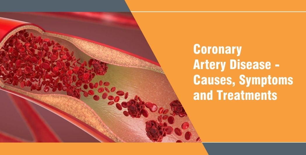
Ageing and other complex factors cause these ordinarily soft and compliant blood vessels to harden. In addition, fat, cholesterol, and minerals from the blood are deposited on the inner surface of the coronary arteries. When this material builds up, it forms a plaque that may restrict the blood flow through the coronary artery. Such plaque may also change the surface of the artery from smooth to rough, and these rough surfaces may stimulate the formation of a blood clot, which may slowly build up and narrow the artery even more. A blood clot can also build up quickly and abruptly close off the artery.
What are the Effects of Coronary Artery Disease?
Narrowed coronary arteries mean the amount of blood that reaches the heart muscle is reduced. Fatigue, tightness in the chest, or a peculiar crushing type of chest pain called angina pectoris may accompany the decreased blood flow. Such symptoms are caused by exercise and emotional stress, which cause the heart to require more blood. However, these symptoms can be handled with adequate rest.
If a coronary artery suddenly closes, blood flow to a part of the heart may stop completely. In this case, some portion of the heart may be permanently damaged. This is often accompanied by severe chest pain that won’t disappear. This is called myocardial infarction, or more commonly, a heart attack. The heart can heal, but the muscle is replaced by scar tissue that doesn’t contract. If the scar is small, recovery can be complete. If the scar is large, it may permanently affect the pumping ability of the heart. It is therefore important that blood supply to the heart be restored before a Heart Attack can occur.
Symptoms of Ischemic Heart Disease
- Chest Pain (Angina) – The word ‘angina’ means ‘strangling’ . The person may feel pressure or tightness in the chest, which could feel similar to strangulation. This pain or tightness usually occurs on the middle or left side of the chest. Angina is generally triggered by physical or emotional stress. The pain usually goes away within minutes after the stressful activity is stopped. In some people, especially women, the pain may be brief, sharp, and felt in the neck, arm, or back.
- Shortness of breath – Shortness of breath happens when a person cannot get enough air into the lungs. The person could struggle to draw a full breath. It might feel like they have just run a sprint or climbed several flights of stairs.
- Heart attack – A completely blocked coronary artery can cause a heart attack. The symptoms of a heart attack (myocardial infarction) are similar to those of angina. Yet, they are not the same. While angina is caused by a drop in blood supply to the heart, a heart attack happens when there is a sudden lack of blood supply to the heart muscle. This blockage is often due to the buildup of plaque in the artery. A heart attack is often accompanied by sweating, fatigue, dizziness, nausea, and vomiting.
How is Coronary Artery Disease Diagnosed?
Treatment for Ischemic heart disease begins with seeking medical care from a healthcare provider. To determine whether or not a person has ischemic heart disease, the health care provider will ask them to undergo several diagnostic tests.
- Electrocardiogram (ECG) – An electrocardiogram records electrical signals as they travel through the heart. An ECG can often reveal evidence of a previous heart attack or one that’s in progress.
- Echocardiogram – An echocardiogram uses sound waves to produce images of the heart. During an echocardiogram, the doctor can determine whether all parts of the heart wall are contributing well to the heart’s pumping activity. Parts of the heart’s function may have been damaged during a heart attack, or parts of the heart might be receiving too little oxygen. This may be a sign of coronary artery disease (Ischemic Heart Disease).
- Exercise stress test – If the signs and symptoms occur most often during exercise, doctors may ask the patients to walk on a treadmill or ride a stationary bike during an ECG. Sometimes, an echocardiogram is also done while the patient exercises. This is called a ‘Stress Echo Test’. In some cases, medication could be used to stimulate the heart instead of exercise.
- Nuclear stress test – This test is similar to an exercise stress test but adds images to the ECG recordings. It measures blood flow to the heart muscle at rest and during stress. A tracer is injected into the bloodstream, and special cameras are used to detect areas that are provided with less blood flow.
- Cardiac catheterization and angiogram – During cardiac catheterization, a doctor gently inserts a catheter into an artery or vein in the groin, neck, or arm and up to the heart. X-rays are used to guide the catheter to the correct position. Sometimes, a dye is injected through the catheter. The dye helps blood vessels show up better on the images and outlines any blockages.
- Cardiac CT scan – A computerized tomography (CT) scan of the heart can help the specialist see calcium deposits in the arteries that can limit blood flow. If a significant amount of calcium is discovered, Ischemic heart disease might be probable. In a CT Coronary Angiogram, the patient receives a contrast dye, given intravenously during a CT scan. This allows detailed pictures of the heart arteries to be captured.
What Can be Done to Relieve Blockage in the Coronary Arteries?
Plaques that block the coronary arteries usually occur in localised portions of the arteries. The part of the artery beyond the narrowing or closure is often not blocked. When the disease is localised in one or two arteries, the blockage can sometimes be opened by stretching or dilating. This is done by using a small balloon on a tube inside the artery. This procedure is called Angioplasty or PTCA (Percutaneous Transluminal Coronary Angioplasty).
Angioplasty has become a relatively common procedure in recent years. Patients around the world will testify to its success in their lives. That said, it still has a few risks attached.
Your doctor will discuss the risks that specifically relate to your condition.
How is Angioplasty Performed?
Patients who have undergone an Angiogram will find. The angioplasty procedure is almost similar. Angioplasty is also performed in a Cardiac Cath lab; Patients usually receive medication before and during the angioplasty procedure to help them relax. You are awake and alert throughout the procedure and are required to respond to the doctor’s requests during the procedure.
Angioplasty begins by inserting a sheath for the catheter into a blood vessel, most often in the upper leg or groin area, but sometimes in the arm.
A very small balloon catheter is passed through the sheath and into the blood vessel leading to the coronary arteries.
With the help of the X-ray, the Cardiologist follows the path of the catheter on the fluoroscope. Pictures may be taken just like in an angiogram.
Once the balloon is at the narrowing of the artery, the balloon is centered, it is then inflated to open the blockage.
While every situation is unique, inflation in most cases will last from 30 seconds to several minutes, depending on the nature of the blockage. The balloon is inflated at least two times. However, it may also be inflated to ten or more times.
While the balloon is inflated, some people experience chest pain that is similar to the angina they have experienced. This happens because the balloon temporarily blocks off the flow of blood and the oxygen that it carries to the heart. Patients should report any pain they feel during the procedure to the Doctor. After the block is opened, the balloon is deflated and retracted back through the blood vessel.
Can Angioplasty be Performed in Other Arteries?
Balloon Angioplasty has expanded the scope of treatment from patients of Coronary Artery Disease to those suffering from blockages in the other arteries of the body. The procedure is being increasingly used to open blockages in the Carotid artery, Renal artery, and other Peripheral arteries.
What is a Stent?
A stent is an expandable mesh tube of a special metal that is designed in a cylindrical wave pattern. The shape and material of the stent offer flexibility for a balloon to adapt to the shape and curves of the artery.
A Stent is implanted to support the artery and keep the vessel open like a structural framework. It is introduced into your artery just after balloon Angioplasty and is positioned at the site of the obstruction. The implantation of a stent benefits those patients who have a potential for future problems, such as block recurrence or restenosis. The stent is implanted permanently in the artery.
What is Percutaneous Transluminal Rotational Ablation (PTCRA)?
One of the treatment options for Coronary Artery Stenosis is the PTCRA or Rotablator System, which is used as a stand-alone treatment or in conjunction with PTCA.
The Rotablator system is a cather-based Angioplasty device utilizing a diamond-coated elliptical burr at the tip of a flexible drive shaft. Tracking coaxially over a guide wire and rotating at upon 190,000 revolutions per minute, the burr preferentially cuts away plaque while avoiding healthy tissue. The burr ablates plaque into fine particles that are disposed off by the body’s reticuloendothelial system.
What Happens After the Angioplasty Procedure?
After the Angioplasty procedure, patients are taken to an Intensive Care Unit with special monitoring equipment. Blood pressure, pulse monitoring, and ECGs are performed routinely after Angioplasty procedures and do not signify any special problems. Suppose a patient experiences any chest discomfort or pressure. The nurse should be notified immediately.
Recovery in the hospital is most likely a matter of allowing the insertion site to heal before getting up and walking around. Most patients are required to hold their leg or arm straight and still for the first six to eight hours. You may be required to stay in the Hospital for 3-5 days before being discharged to the care of your family doctor. You can resume full activity within a few days of returning home.
Angioplasty is not a cure, but a treatment to reduce the effects of Coronary Artery Disease. It is extremely important to follow your medication regimen without deviation. Another element for quick recovery involves accommodating lifestyle changes to improve their health and minimizing the impact of their heart disease. Several factors are known to contribute to the buildup of plaque in the coronary arteries. It is the combination of several of these risk factors, rather than a single factor, that impacts coronary artery disease.
Some of these risk factors such as male sex, age and heredity can only be attended but cannot be changed. However, other factors that can be controlled include:
- High Blood Pressure
- Smoking
- High Blood Cholesterol
- Body Weight
- Diabetes
- Lack of Proper Exercise
- Stress
What Can a Patient Do to Manage Ischemic Heart Disease?
- Keeping the blood pressure, cholesterol, and blood sugar levels in check
- Cutting back on alcohol
- Quitting the use of tobacco products
- Managing the stress
- Practicing regular physical activity
Coronary heart disease (Ischemic Heart Disease) cannot be cured, but treatment can help manage the symptoms and reduce the chances of medical emergencies such as a heart attack.


