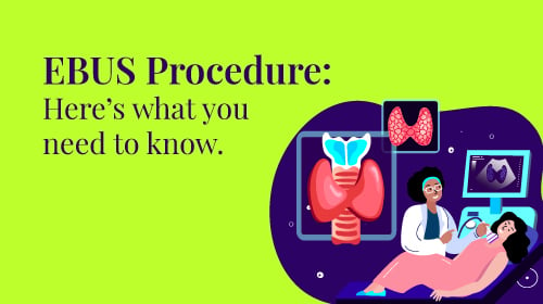
All About EBUS Procedure
August 17, 2022

Overview
An Endobronchial ultrasound (EBUS) allows your doctor to see the airways that lead to your bronchi. Additionally, it might assist your doctor with a treatment like a biopsy.
You won’t need to have any surgical cuts made to your chest for an EBUS because a bronchoscope is used to perform this task. However, if your doctor suspects you have a lung ailment such as an infection, lung cancer, lymphoma, or an inflammatory illness like sarcoidosis, they may order an EBUS.
Lung cancer can be diagnosed or its stage determined using endobronchial ultrasound (EBUS), a medical procedure carried out during a bronchoscopy. A flexible scope is inserted through the mouth into the major bronchi of the lungs during EBUS to scan tissues utilising high-frequency sound waves.
Endobronchial ultrasonography is risk-free and minimally invasive, requiring neither surgery nor ionising radiation exposure. It can also aid in diagnosing some inflammatory lung disorders that cannot be validated by conventional imaging testing. It is often done as an outpatient procedure.
Purpose of the Procedure
Endobronchial ultrasonography may be prescribed in addition to conventional bronchoscopy if you have been diagnosed with lung cancer or if your initial tests strongly show that you may have it.
Contrary to bronchoscopy, which uses a viewing scope to observe the airways directly, EBUS uses refracted sound waves to allow medical professionals to look through the airways’ tissues.
Endobronchial ultrasound can be used to assess the degree of tumour invasion in the central airways, which may happen in cases of metastatic lung adenocarcinomas and squamous cell carcinomas (which generally start in the airways) (which can spread from the outer edges of the lungs and invade the central lung).
The following are the two main EBUS indications:
- Staging of Lung cancer: To ensure that the right treatment is given, lung cancer staging is used to assess the severity of the disease. Using a transbronchial needle aspiration procedure, endobronchial ultrasonography enables medical professionals to take tissue from the lung or mediastinal lymph nodes in the chest (TBNA). The biopsied cells can then be sent to the lab for study to identify how early or advanced the cancer may be.
- Evaluation of abnormal lesions: If a chest X-ray or computed tomography (CT) scan reveals an abnormal lesion, EBUS with TBNA can be utilised to collect a sample of the afflicted tissues. This can assist in determining whether cancer or an inflammatory lung disease like sarcoidosis is to blame for swollen lymph nodes. In addition, pulmonary lymphoma, a type of blood cancer, can be suspected in persons with lymph nodes that can be sampled using EBUS.
Typically, a doctor won’t use endobronchial ultrasonography as their initial method of a lung cancer diagnosis. However, it is often requested when preliminary lab and imaging testing reveals the condition’s presence. With EBUS, your healthcare professional can access a mass or nodule close to a significant airway without cutting through the chest wall.
Before the Procedure
Endobronchial ultrasonography, like bronchoscopy, is generally considered safe with a low risk of complications. Preparing is easier when you know what to expect.
Timing
Even though the EBUS technique only takes 20 to 30 minutes to complete, getting ready for the surgery and recovering from the anaesthetic can take up to four hours. Therefore, it is recommended to cancel all of your appointments on the day of your treatment and plans a second day off so you can recover.
What to Wear
Dress comfortably with items of clothing that are simple to take off and put back on because you will be required to change into a hospital gown. Leave any jewellery or valuables at home if at all possible.
Before the operation, be ready to take off any dentures, hearing aids, contacts, or eyeglasses. You’ll be provided with a safe location to store items in addition to your wardrobe and other possessions.
Food and beverage
You’ll typically be instructed to stop eating at midnight the day before the surgery. Most EBUS procedures are scheduled for the morning to prevent you from being overly hungry. The healthcare professional could urge you to cease eating six hours before the procedure if it is planned for later in the day.
During the EBUS Procedure
The bronchoscope is a tiny, illuminated tube inserted by your doctor into your mouth, down your windpipe, and into the bronchi. Your doctor can view your airways, blood vessels, lungs, and lymph nodes on an ultrasound monitor thanks to a tiny camera attached to the bronchoscope.
To collect tissue and fluid samples from your lungs and nearby lymph nodes, your doctor can use the bronchoscope, which also contains a fine needle. Transbronchial needle aspiration is the procedure’s name, and biopsy is the name of the sample.
Following EBUS After the surgery, you won’t be able to cough for a few hours. For a few days, your throat could feel sore and scratchy.
After the procedure
Your doctor will send the biopsy to a laboratory for analysis. Your doctor can determine the stage of your lung cancer using the information from the needle aspiration.
Your doctor will inform you of the findings and any potential future measures.
People also ask
1. Why is EBUS done?
An EBUS (endobronchial ultrasound) bronchoscopy is used to identify various lung conditions, such as inflammation, infections, or malign.
2. Is EBUS biopsy painful?
After that, tissue samples are taken from the lymph nodes using a needle. Some patients may report a sore throat following the operation. Next, a pulmonologist carries out EBUS bronchoscopy.
3. How long does an EBUS procedure take?
The length of an EBUS procedure varies depending on the results and whether therapy is required, although it often lasts 45 minutes or less. You should plan to spend two to four hours in the department, not counting the time spent getting ready for the treatment and recovering.
4. Is an EBUS a biopsy?
Lung cancer, infections, and other disorders affecting enlarged lymph nodes in the chest can all be identified with endobronchial ultrasonography (EBUS), a minimally invasive but extremely efficient method.


