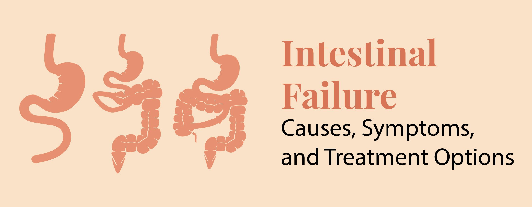
Robotic Surgery at Rela Hospital Repurposes Small Intestine into Bladder and Eliminates Patient’s 10-Year Long Dependence on Urinary Catheter
November 6, 2023
- Ileocystoplasty, and appendicovesicostomy, both urological procedures, are done simultaneously using robotic assistance
- Robot-assisted surgery comes as a boon for patients who have dysfunctional bladder, as it makes it possible to create or augment a bladder using a segment of the small intestine, all without the need for long incisions
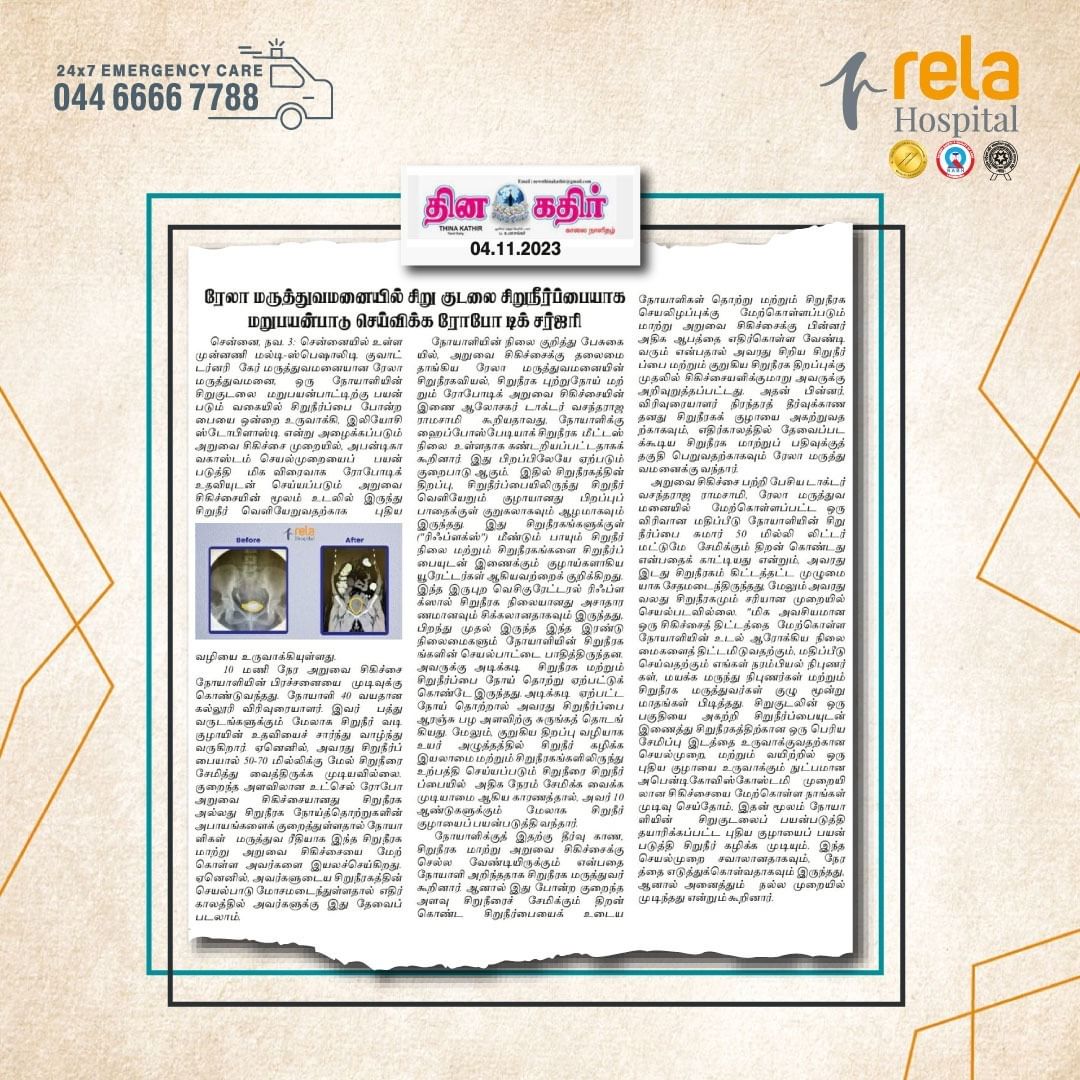
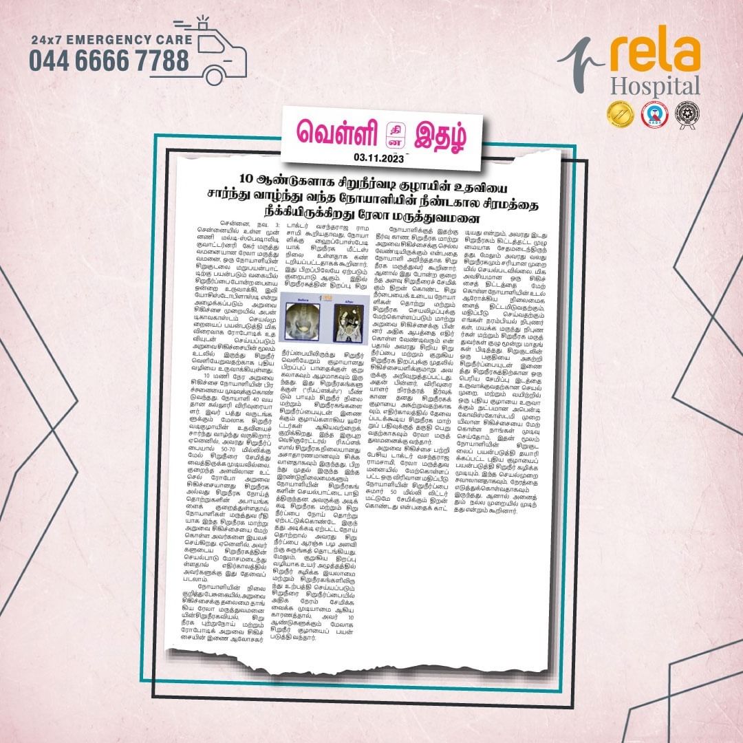
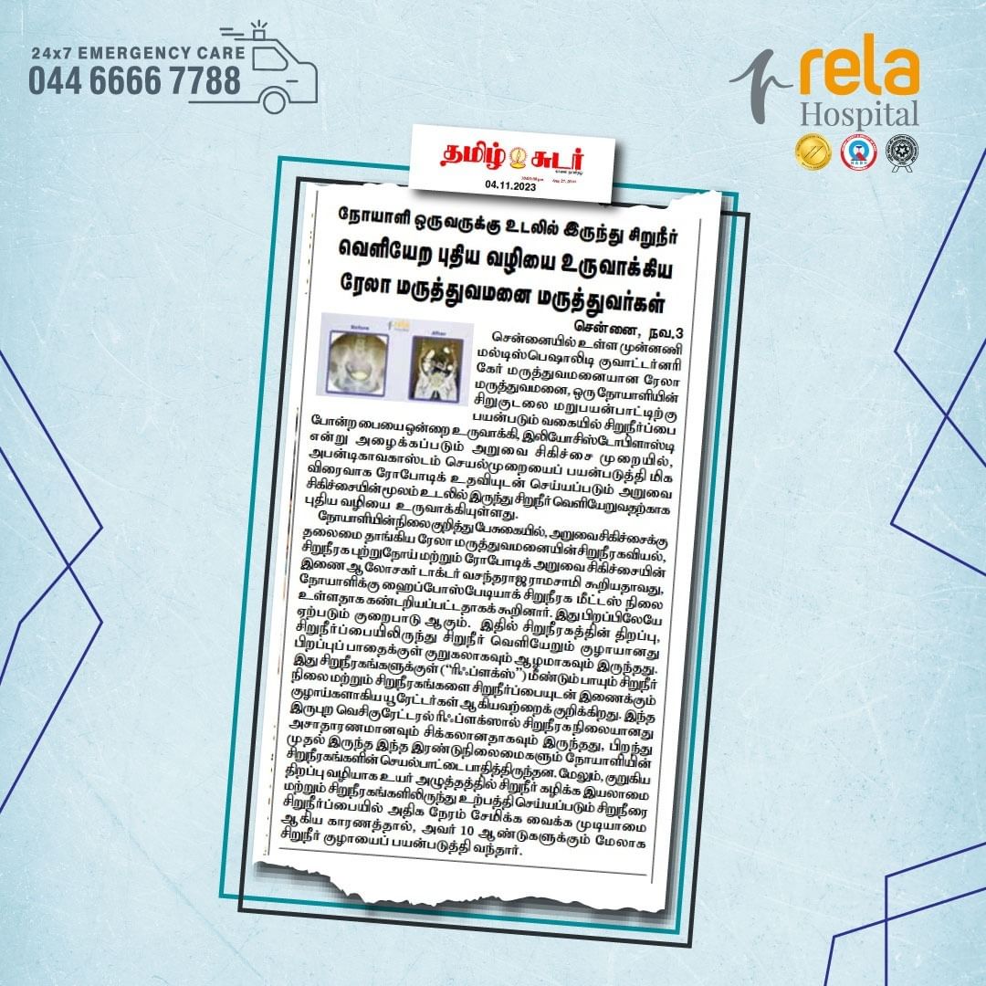
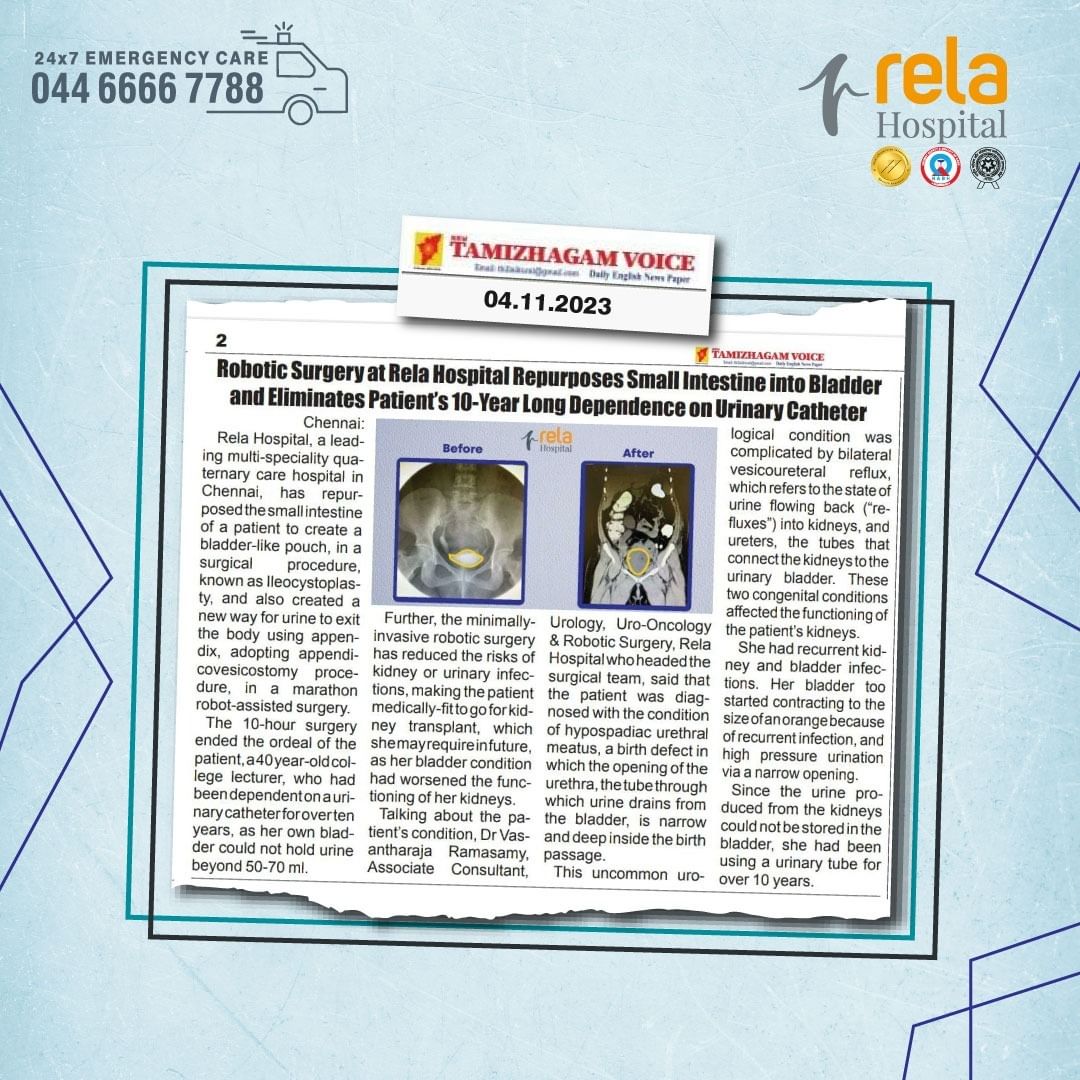
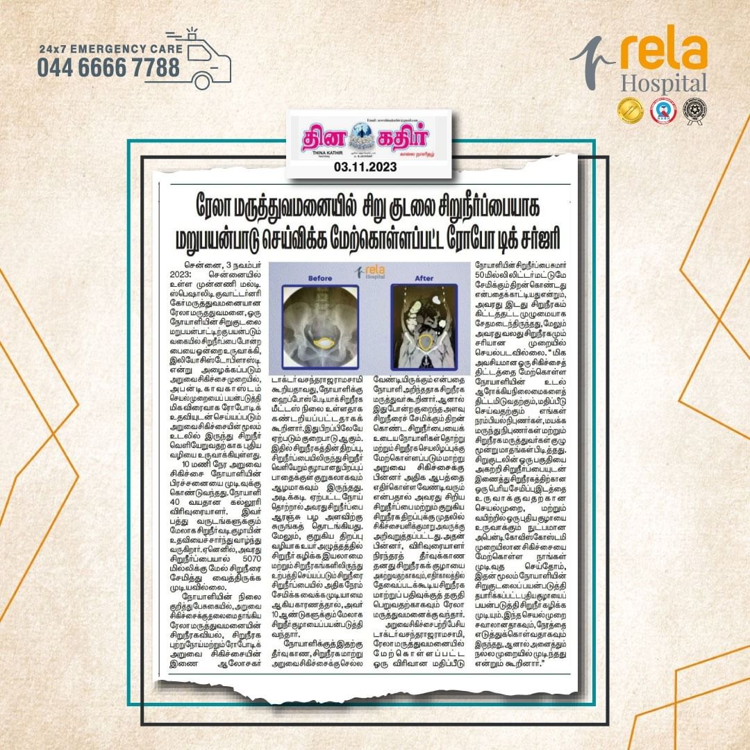
Chennai, 31 October 2023: Rela Hospital, a leading multi-speciality quaternary care hospital in Chennai, has repurposed the small intestine of a patient to create a bladder-like pouch, in a surgical procedure, known as Ileocystoplasty, and also created a new way for urine to exit the body using appendix, adopting appendicovesicostomy procedure, in a marathon robot-assisted surgery.
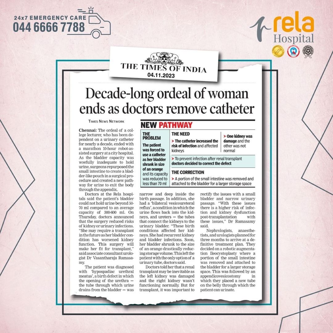
The 10-hour surgery ended the ordeal of the patient, a 40 year-old college lecturer, who had been dependent on a urinary catheter for over ten years, as her own bladder could not hold urine beyond 50-70 ml. Further, the minimally-invasive robotic surgery has reduced the risks of kidney or urinary infections, making the patient medically-fit to go for kidney transplant, which she may require in future, as her bladder condition had worsened the functioning of her kidneys.
Talking about the patient’s condition, Dr Vasantharaja Ramasamy, Associate Consultant, Urology, Uro-Oncology & Robotic Surgery, Rela Hospital who headed the surgical team, said that the patient was diagnosed with the condition of hypospadiac urethral meatus, a birth defect in which the opening of the urethra, the tube through which urine drains from the bladder, is narrow and deep inside the birth passage. This uncommon urological condition was complicated by bilateral vesicoureteral reflux, which refers to the state of urine flowing back (“refluxes”) into kidneys, and ureters, the tubes that connect the kidneys to the urinary bladder. These two congenital conditions affected the functioning of the patient’s kidneys. She had recurrent kidney and bladder infections. Her bladder too started contracting to the size of an orange because of recurrent infection, and high pressure urination via a narrow opening. Since the urine produced from the kidneys could not be stored in the bladder, she had been using a urinary tube for over 10 years.
On the solution the patient needed, the urologist said that the patient came to know that she might have to go for a kidney transplant. But she was advised to get treated for her small bladder and narrow urinary opening first, as patients with such limited bladder capacity face a higher risk of infection and kidney dysfunction post-transplantation. The lecturer then came to Rela hospital for the permanent solution of getting rid of her urinary catheter as well as for making her eligible for kidney transplant registration which may be required in future.
Talking about the surgical intervention, Dr Vasantharaja Ramasamy, said that a comprehensive assessment at Rela Hospital showed that the patient’s bladder had a capacity of only about 50 millilitres. Her left kidney was almost completely damaged, and her right kidney too was not functioning optimally. “Our team of nephrologists, anaesthetists, and urologists, took three months for planning and evaluation of the patient’s health conditions, to arrive at a definitive treatment plan. We decided to do a robotic augmentation Ileocystoplasty, the procedure to remove a portion of the small intestine and attach it to the bladder to create a larger storage space for urine, and to do appendicovesicostomy, a technique to create a new tube on the belly through which the patient can urinate by using a new tube made using the appendix. The intervention was challenging and time consuming but it went off well.”
The patient’s small intestine was repurposed to create a new, larger bladder-like pouch with a volume of 300-400 ml. This pouch was sutured to the patient’s native bladder. The patient’s own appendix was employed as a conduit and sutured to the abdominal skin, ensuring a permanent urinary opening. The entire procedure was performed robotically, reducing the need for a long abdominal incision and utilising only four small 8 mm incisions in different quadrants of the abdomen.
Following these rare and complex procedures, the patient is freed from the urinary catheter. She also learned to manage self-urinary drainage through the small opening in her abdomen. This transformation not only improved her quality of life but also made her an eligible candidate for potential kidney transplantation.

