Infrastructure, Technology & Other Diagnostic Procedures
Besides high-end surgical techniques, the department also offers a wide range of diagnostic procedures in order to decide upon the further course of treatment.
Electrocardiography (ECG)
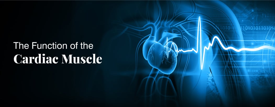
An electrocardiography or ECG helps in measuring the electric activity of the heartbeat. The ECG will analyse the timing of top and lower chambers. An abnormal ECG is usually an indication of impending cardiac problem and would help the cardiologist to decide upon the further course of treatment.
Treadmill Test
Treadmill test or a stress test assess how your heart is functioning during intense activity like exercise, work. The test reveals if the blood supply is getting reduced while the patient is running on the treadmill and if the heart is pumping more blood. It measures heart rate, blood pressure, ECG and the doctor will also consider how tired the patient is after the test.
3D Echocardiography
The 3D Echocardiography or 3Dimensional Echocardiography gives complete evaluation of both left ventricle and right ventricle, ejection fraction with imaging reference. Since it provide the total picture of the functioning of heart, it helps the cardiologist in reaching an accurate diagnosis.
Advanced Echocardiography
An advanced echocardiography provides advanced imaging to diagnose the conditions like atrial and ventricular septal defects, blocked heart valves and various other abnormalities related to heart functioning. With the help of clear imaging, the cardiologist will understand the condition of the patient and decide upon the further treatment.
Transoesophageal ECHO Cardiography
Transesophageal Echo Cardiography or TEE is a test that provides with complete imaging of the heart functioning aided by ultrasound. Under this procedure, a thin tube is passed through the mouth and then esophagus. Since the esophagus is closely located to heart if facilitates better pictures with utmost clarity to understand the structure of the heart and valves.
128 Slice Cardiac CT
128 Slice Cardiac Computing Tomography provides overall information about the heart and its functioning by providing the cardiologists all necessary information to rule out or diagnose life threatening conditions like pulmonary embolism, aortic dissection, acute coronary syndrome. The test provides computerized imaging procured through narrow beam of x-rays even in the cross-sectional or slices as they are often referred to.
Holter Monitoring
Holter monitoring is done to evaluate the heart’s activity for a period of 24 hours to 48 hours. A battery operated portable device, it is attached to the skin with electrodes. Doctors would recommend holter monitoring if the patient complains of fast or slow heartrate, feeling dizzy and also to assess the functioning of pacemaker.
Myocardial Perfusion Scintigraphy
Myocardial Perfusion Scintigraphy is a nuclear medicine imaging test in which radioactive tracer gets injected in small amounts to assess the blood supply to the heart. It gives a clear understanding of the difference in blood supply during rest and stress and also helps the doctor to diagnose the damage to the heart after suffering a heart attack.
PET-Scan
Positron Emission Tomography or PET scan is a nuclear medicine imaging test in which radioactive tracer gets injected in small amounts to take pictures of the heart. It is recommended in the case of diagnosing coronary artery disease and also to assess the damage of heart following a heart attack. In certain cases, it is also done to assess if an angioplasty, stenting, coronary artery bypass surgery would help the patient in complete recovery.


