Cutting Edge Technologies Fight Brain Tumours into Remission
April 9, 2025

A brain tumour is a mass of abnormal cells in the brain. It can be benign or malignant. A malignant tumor can be primary, originating in the brain, or secondary, where cancers spread to the brain from other parts of the body. The size, location, and rate of growth of a tumour will determine the severity of symptoms and the treatment options.
Symptoms of Brain Tumours
There are many types of brain tumours. The symptoms are common to all and they include:
- New headache or changing pattern of headaches.
- Gradual increase in frequency and severity of headaches.
- Unexplained nausea or vomiting.
- Vision/ Speech/ Hearing problems.
- Gradual loss of arm/ leg sensation or movement.
- Balance problems.
- Confusion.
- Behavioral changes.
- Sudden onset of seizures.
Modern Diagnostic Tests for Brain Tumours
A neurological exam is the first step in diagnosis. A neurologist will examine your vision, hearing, balance, coordination, reflexes, and strength. This will help identify the areas affected by the tumor.
Imaging Tests
Magnetic resonance imaging (MRI) scans, including functional MRI, perfusion MRI, and magnetic resonance spectroscopy, are used to detect and stage the tumour and plan treatment. Computerised tomography (CT) and Positron emission tomography (PET) can be used to check for cancer in other parts of the body if the tumour is suspected to be a secondary tumour.
Stereotactic Biopsy
Tissue is removed from the tumour through regular biopsy or a stereotactic biopsy. Stereotactic biopsy may be done for tumours in hard-to-reach or sensitive areas. A small hole is drilled in the skull, and a thin needle is inserted through it. Tissue is removed using the needle. The process is guided by CT or MRI scanning. The sample is examined to determine if it contains cancerous cells.
Molecular diagnostics, sophisticated laboratory tests, and advanced imaging technologies, such as high-powered (7-tesla) MRI scans and magnetic resonance elastography (MRE), enable doctors to plan individualized treatments. This reduces the need for follow-up surgery.
Advanced Treatment Methods for Brain Tumours
Doctors proceed with the treatment based on the type, size, and location of the tumour. Treatment options include:
Surgery
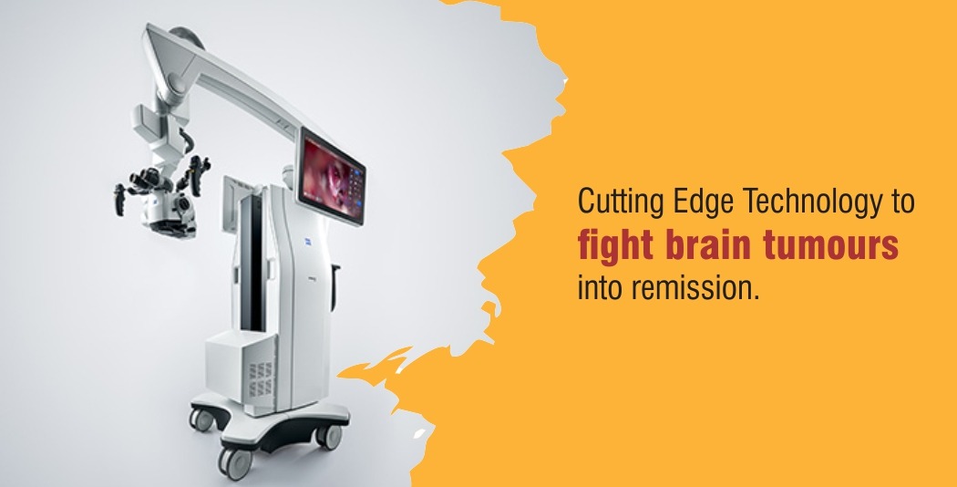
Surgery is performed to remove the tumour or a part of the tumour if it is easily accessible. This will provide relief from symptoms. Neuroradiologists, specialists in brain imaging, utilize advanced surgical navigation and mapping equipment to achieve greater accuracy during complex surgeries.
Advanced surgical procedures include
- Awake brain surgery (Awake craniotomy)
- Minimally invasive surgery
Awake Craniotomy – If a brain tumor that causes seizures needs to be removed, awake craniotomy is performed to ensure the safety of the language, speech, and motor centers of the brain. This requires your active participation, so you have to be awake during a portion of the surgery. The anesthesiologist puts you to sleep when a portion of the skull is removed. He then stops administering the sedative and allows you to wake up. The neurosurgeon will conduct brain mapping to determine the control centers of your brain. During surgery, you will be asked questions, shown pictures to identify, and make simple movements. Your responses will further help the surgeon identify the functional areas of your brain. Based on this and the brain mapping, the surgeon safely removes as much of the tumour as possible while preserving maximum brain function. Once the surgery is complete, the anesthesiologist will put you to sleep, and the surgeon will reattach the skull.
Brain Mapping
The brain is filled with electrical impulses. During brain mapping, these electrical impulses are measured with an Electroencephalogram (EEG). Functional MRI (fMRI) is used to map abnormal blood flow in a specific area of the brain. Positron Emission Tomography (PET) is used to image brain tissue and its activity after administering a radioactive tracer. Brain Mapping is primarily used to identify the main motor, sensory, and visual areas of the brain, thereby minimizing damage during the surgical removal of a brain tumor while maximizing tumor removal.
Radiation Therapy
High-energy beams, such as X-rays or protons, can be used in radiation therapy to kill tumour cells. An external radiation source or a radiation source placed inside the body close to the brain (brachytherapy) may be used.
Radiosurgery
A Gamma knife or linear accelerator is used to deliver radiation therapy. Multiple beams of moderate radiation are focused on a small area to provide a large dose, killing tumor cells.
Chemotherapy
Drugs are given either orally or intravenously to kill tumour cells. The type of brain tumour is determined, and if suitable chemotherapy is used.
Targeted Drug Therapy
Certain drugs can target specific abnormalities present in the cancer cells to kill them. Targeted drug therapy is available for some types of cancers.
While a brain tumour diagnosis can be overwhelming, today’s advanced medical technologies, combined with comprehensive rehabilitation, offer real hope. With timely intervention and continued care, many patients can not only survive but also reclaim a fulfilling and meaningful life beyond cancer.


