Interventional Cardiology
Invasive Diagnostics and Interventions
Primary Angioplasty 24x 7 With A Door to Balloon Time Of 30 -60 Minutes
Primary Angioplasty also known Percutaneous Coronary Intervention is a life-saving procedure which is usually done in the cases of acute myocardial infarction or heart attack in layman’s terms. It treats blocked coronary arteries, and restores immediate blood flow to the heart. Under local anaesthesia, a catheter gets inserted into the main artery through the thigh or arm. The balloon passed over the wire through the catheter then gets inflated opening the artery after which a medicated metal stent gets placed facilitating blood flow. The stent thus becomes the permanent part in the body and reduces the risk for further cardiac events.
Since, every second counts in saving the life during a heart attack, this facility is available 24×7 and we ensure performing it within 30 to 60 minutes, soon after suffering from it.
Elective Coronary Angiography and Angioplasty
An elective Coronary Angiography and Angioplasty is done to diagnose and to treat the conditions related to heart and blood vessels. It is recommended if the patient complains about chest pain, discomfort or pain in jaw, neck and arms, sudden episodes of chest pain, abnormal results in heart stress tests, a heart valve problem, congenital heart disease. The elective angiography and angioplasty serve as preventive measures to save the heart from an impending heart attack or a stroke.
The Coronary Angiogram procedure is aided by an x-ray machine and a dye gets inserted into the blood vessels. The x-ray machine then takes the imaging of blood vessels and the expert would be able to view clogged arteries, if any. These images are called angiograms.
If the expert diagnoses clogged arteries, then an angioplasty would follow the angiogram. Angioplasty or Percutaneous Coronary Intervention is done under the local anaesthesia, in which a catheter gets inserted into the main artery through the thigh or arm. The balloon passed over the wire through the catheter then gets inflated opening the artery after which a medicated metal stent gets placed facilitating blood flow. The stent thus becomes the permanent part in the body and reduces the risk for further cardiac events.
Complex Coronary Angioplasty with Advanced Imaging Technology (IVUS & OCT)The cardiac procedures have undergone major technological advancements in the recent years helping the medical fraternity in much faster and accurate diagnosis. Complex Coronary Angioplasty with Imaging Technology is one such invention that is greatly aiding the medical experts.
IVUS: Known as Intravascular Ultrasound, IVUS is usually recommended to determine the amount of plaque build-up in epicardial coronary artery. A specially designed catheter with a tiny ultrasound probe attached to computerized ultrasound equipment gets inserted into the coronary arteries. The IVUS not only provides a complete understanding of the regression and progression of artherosclerotic lesions but also helps the expert take stock of hidden plaque or atheroma. This procedure comes in diagnosing patients with restenosis.
Intravascular OCT:
Intravascular Optical Coherence Tomography helps in examining coronary arteries and this procedure offers high resolution of the blocked tubes. It can in fact help the expert doctor in differentiating between fibrous, lipid-rich or calcified plaque. It also provides other detailed information during angioplasty helping the cardiologist to decide upon the type of stent implantation especially in the patients suffering from restenosis.
Chronic Total Occlusion PCI
A Chronic Total Occlusion Percutaneous Coronary Intervention is done to clear blockages that have been present for more than 3 months, often a result of atherosclerosis. It is done in the case of completely blocked arteries and the duration of the procedure varies from patient to patient. This requires a greater expertise as the doctor uses both antegrade and retrograde approaches to reach to the blockage. After identifying the blockage, the expert cardiologist would insert catheter into the collateral vessels for entering the blocked artery. An inflated balloon then gets placed and blown up at the tip of the catheter facilitating a wide opening of the arteries for restoring blood flow and also place a stent.
FFR Guided PCI
Fractional Flow Reserve Percutaneous Coronary Intervention helps in ascertaining the pressure differences in case of coronary artery stenosis. It gives a fair idea to the cardiologist if the stenosis is blocking oxygen delivery to the heart muscle, a condition known as myocardial ischemia. During the procedure, a catheter gets inserted through the groin and an FFR – a small sensor at the tip of the catheter measures the pressure, temperature and severity of the lesion. This procedure helps the expert doctor to determine the amount of pressure decline caused due to vessel narrowing.
Diagnostic Cardiac Catheterisation
Cardiac Catheterisation is a diagnostic procedure to treat various cardiovascular conditions. Under this procedure a long, thin catheter gets inserted into the body via groin, arm or neck to locate narrowed or blocked arteries, measure oxygen and pressure, to check the functioning of right and left ventricles, to extract a tissue from the heart, diagnosing congenital heart ailments and also to study defects in heart valves.
It is also often used as a part of angiogram and angioplasty for interventional purposes.
Percutaneous Valvuloplasty
A Percutaneous Valvuloplasty is a technique where a single or multiple balloon are inserted beneath the skin and then get inflated to open a stenotic valve. It is recommended for patients suffering from mitral, aortic and pulmonic stenosis. It serves as an amazing option for those patients at the high risk of surgical complications and it provides an immediate and a great degree of relief from symptom
Transcutaneous Aortic Valve Implantation (TAVR)
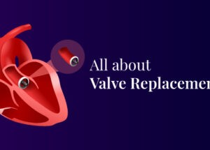
Transcutaneous Aortic Valve Implantation or TAVR is done when the aortic valve fails to open and is a great option for the patients at the high risk of surgical complications following an open-heart surgery. Patients with kidney damage and lung related ailments too get benefited through this procedure. It will decrease major symptoms like recurrent chest pain, fainting, swelling in the legs and prevents sudden cardiac failure and death.
During the procedure the surgeon will insert a catheter to access the heart and valve. Soon after positioning the new valve, the balloon gets inflated and replaces the valve in the appropriate place. The catheter is removed after securely placing the valve.
Implantation of Pacemakers
Pacemaker is a small electronic device that is placed in the chest for regulating slow electric problems in the heartbeat. Comprised of three parts – a pulse generator, one or more leads and an electrode on each lode, pacemaker sends signals to the heart to beat when it going dangerously low or going irregular. It is needed for the patients who suffer from the lack of electric stimulation of the heart to heart muscle and the response from heart’s chambers.
Pacemaker implantation is done under local anaesthesia. A small incision will be under the collarbone and the pacer will be slowly placed with the help of lead wire into the blood vessel and then to the heart. After it reaches inside the heart, the functioning will be tested aided by fluoroscopy and ECG. The pacemaker generator will be then placed on the non-dominant side of body (if you are right-handed person it is placed on to your left and vice-versa). The patient would be able to resume normal duties within few days.
Implantation of Automatic Cardiac Defibrillators
Automatic Cardiac Defibrillators are a boon to patients with a known ventricular tachycarida or fibrillation. The Cardiac Defibrillator plays a crucial role in preventing sudden cardiac arrest in high risk patients. An ICD in few cases even serves as a pacemaker. It is in fact a battery powered device that gets implanted under the skin which monitors heart rate. The moment it recognises an abnormal rhythm it sends electric shock and restores normal heart rate.
It is implanted under the collarbone just below the skin. The leads are attached to the pulse generator and it prevents the need of open-heart surgery.
Implantation of IVC Filters
Inferior Vena Cava Filter of IVC filter is a medical device, implanted into the inferior vena cava a large vein that carries deoxygenated blood to prevent pulmonary emboli. It is implanted a thin catheter is inserted in the inferior vena cava aided by an X-ray or an ultrasound. The filter at the tip of the catheter is then placed and will be securely attached. The catheter is then removed. The filter placed in vena cava traps and stops the blood clots in the lower body, preventing them from travelling to the lungs, thus saving the patient from a life-threatening condition.


